|
What is Raman? How does it work?
 Raman
spectroscopy takes advantage of the inelastic scattering of monochromatic laser
light by molecules. Energy from the laser is exchanged with the molecules in
such a way that the scattered light photons have higher or lower energy
than the incident photons. The difference in energy is due to a change in the
polarization energy of the molecule and gives information about
the molecular structure. Since different molecules show different energy
changes, the Raman technique can be used as a qualitative analysis method. Raman
spectroscopy takes advantage of the inelastic scattering of monochromatic laser
light by molecules. Energy from the laser is exchanged with the molecules in
such a way that the scattered light photons have higher or lower energy
than the incident photons. The difference in energy is due to a change in the
polarization energy of the molecule and gives information about
the molecular structure. Since different molecules show different energy
changes, the Raman technique can be used as a qualitative analysis method.
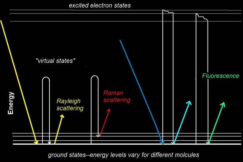
Diagram of Raman scattering: Incident light (yellow) that loses or gains
no energy is scattered back at the same wavelength is called Rayleigh
scattering. If some of the energy is transferred to the ground state, the
scattered light is scattered at a longer wavelength (red). Fluorescence is
another effect that causes light to be re-emitted at longer wavelengths.
It often masks Raman scattering.

Diagram of Raman instrumentation: Incident laser light (yellow) is
scattered at the light surface. Most of the light is scattered at the same
wavelength at the incident light. Light that is Raman shifted also is
scattered in a random directions. A lens is used to collect the light, and
a filter is used to block the wavelength of the incident light. Longer
wavelengths (Raman scattering) is transmitted to the monochromator and detection
system. The frequency shift of the scattered light will determine the
chemical structure of the sample material.
Symphotic TII Raman
Products:
The
Nanofinder® Confocal Microspectroscopy System:
 Announcing the NEW
Nanofinder 30 :
High resolution, high sensitivity, full automation. Announcing the NEW
Nanofinder 30 :
High resolution, high sensitivity, full automation.
A High Sensitivity/High Resolution Scanning Confocal
Raman Spectroscopic Microscope
Our new confocal microscope, the “Nanofinder
30”, is designed for simultaneous high spatial
resolution (200 nm) and sensitivity (to single photon counting). This system is
fully automated and features a new multi-laser excitation capability. While
normal confocal microscopy permits observations in three spatial dimensions,
this new instrument is equipped with a spectrometer and time correlated single
photon counter, adding two more analytical dimensions: spectral and temporal.
Designed for low level signal detection, the “Nanofinder
30”has
applications in 3D Raman and photoluminescence imaging, single molecule
fluorescence detection, Raman spectral and spatial analysis of a variety of
materials such as semiconductors and semiconductor devices, CVD artificial
diamond arrays, carbon nanotubes and living cells.
Click
here for single page flyer.
The new
InSITEtm
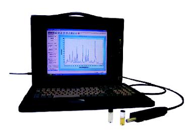 Remote
Probe Raman Spectroscopy System for
in situ Chemical Analysis Remote
Probe Raman Spectroscopy System for
in situ Chemical Analysis
The InSITE Raman probe may be deployed manually with a pole or
manipulator, or on a robot crawler complete with camera for visual
inspection.
Special lenses are available for determining Raman
spectra without contact.
Three example spectra
from materials commonly found in nuclear power plants are shown on the below.
The broad peak shown in the paint spectrum is caused by fluorescence, which is
also measured by the InSITE system, and is also useful in identifying unknown
substances. For a full presentation on the application of the InSITE in
nuclear power plant inspections,
click
here.
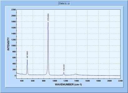
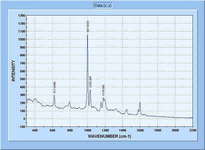
Raman Spectrum of
boric acid deposit on the wall of a power plant.
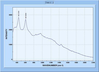
Raman and
fluorescence spectrum of epoxy paint chip from power plant wall
Raman spectrum of
polypropylene plastic chip.
The InSITE System uses a remote Raman probe to non-destructively test in
hazardous areas for chemical analysis of substances such as boric acid,
explosives, or other molecular compounds.
Click here for a brochure.
|


